Research Program Overview
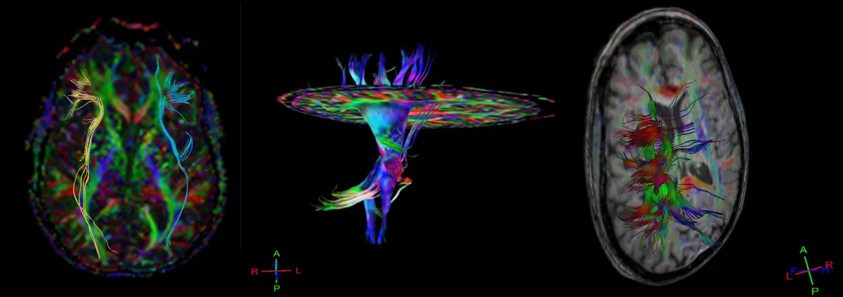
Working with Sheng-Kwei Song, PhD, we are employing Diffusion Basis Spectrum Imaging (DBSI) to detect spinal cord injuries that are not visible with traditional imaging methods. DBSI offers a detailed view of the spinal cord’s internal structure, identifying tissue damage and inflammation types. This technique is crucial for uncovering hidden injuries, providing critical information for early intervention, and potentially improving patient outcomes.
DBSI allows imaging with higher specificity for injury compared to conventional MRI and DTI techniques. Our team is currently validating prior research efforts conducted in mice to humans, building upon the foundational work conducted in the Song Lab. This advanced imaging approach represents a significant advancement in our ability to detect and characterize occult neurological injuries in spine trauma patients.
Clinical Translation
This technique is crucial for uncovering hidden injuries, providing critical information for early intervention, and potentially improving patient outcomes. Our research focuses on translating advanced DBSI imaging techniques from research settings into clinical practice to enhance diagnostic capabilities for spine injury patients.
Research Inquiries Collaboration Opportunities
Mission
To employ Diffusion Basis Spectrum Imaging (DBSI) to detect spinal cord injuries that are not visible with traditional imaging methods, providing critical information for early intervention and potentially improving patient outcomes through advanced neuroimaging techniques.
Primary Research Areas
Occult Injury Detection
Advanced diffusion MRI techniques for detecting spinal cord injuries not visible with traditional imaging methods
Tissue Characterization
Detailed analysis of tissue damage and inflammation type identification using DBSI metrics
Early Intervention
Development of biomarkers for early intervention and improved patient outcome prediction
Comparative Analysis
Comparative imaging analysis between DBSI and conventional DTI techniques for enhanced specificity
Translational Research
Translation of proven mouse model techniques to human clinical applications
Fraction Analysis
Comprehensive analysis of non-restricted, restricted, and fiber fractions in tissue microstructure
Research Methodology
Our research program utilizes state-of-the-art DBSI imaging protocols to quantify different tissue components including non-restricted, restricted, and fiber fractions. We employ advanced image processing techniques to analyze DBSI metrics including apparent diffusion coefficients and fractional anisotropy measurements that provide detailed insights into tissue microstructure. The methodology includes comprehensive validation studies comparing DBSI findings with conventional MRI and DTI imaging, histological correlation studies, and longitudinal tracking of injury progression. We collaborate with the Song Lab to translate proven mouse model techniques to human clinical applications, ensuring robust scientific validation of our imaging protocols.
Current Initiatives
Human Translation Study
Ongoing Validation of DBSI techniques previously proven in mouse models for detection of occult spinal cord injuries in human patients with spine trauma.
Longitudinal Injury Tracking
In Development Development of DBSI protocols to monitor injury progression and healing over time, enabling better prediction of patient outcomes and treatment response.
Clinical Implementation Program
Planning Phase Integration of DBSI imaging into routine clinical workflows for spine trauma patients to improve diagnostic accuracy and treatment planning.